Interesting Oral Pathology Case
A 65 year old man came into my clinic with chief complaint of swelling to the right side of the face. The swelling has been present for the past 2 weeks.
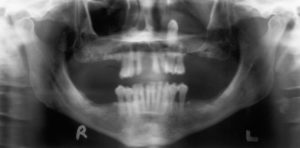
Examination reveals swelling and fluctuance in the left buccal maxillary vestibule. Panoramic image reveals a retained, unerupted maxillary left canine #11, located in the maxillary sinus.
CT scan reveals a large lesion associated with impacted maxillary canine, and eroding through the anterior maxillary sinus wall.
Differential diagnosis in this case (cystic lesion associated with an impacted tooth) would include a dentigerous cyst, odontogenic keratocyst, or an ameloblastoma.
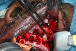
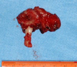
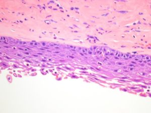
Patient underwent excisional biopsy of the lesion and removal of the impacted tooth, all through an intra-oral approach. Specimen was submitted for pathologic diagnosis. The diagnosis came back as Dentigerous cyst.
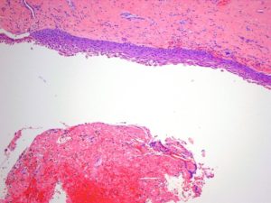
Dentigerous cyst is second-most common odontogenic cyst, with periapical cysts being most common. The cyst develops from proliferation of the enamel organ remnant or reduced enamel epithelium. It is most commonly associated with impacted 3rd molars and maxillary canines (like in this case). The cyst can achieve significant size and can predispose to jaw fracture. The most common appearance of the cyst is unilocular, although multilocular dentigerous cysts can also be seen. Histopathologically, the fibrous connective tissue wall of the cyst is lined by stratified squamous epithelium. In most cases, removal of the associated tooth and enucleation of the cyst is definitive treatment. Under very rare circumstances, the lining of untreated dentigerous cyst can undergo a transformation to an ameloblastoma.
Dr. Dmitry Tsvetov
Comments are closed.


