
Another unusual pathology case
43 year-old female 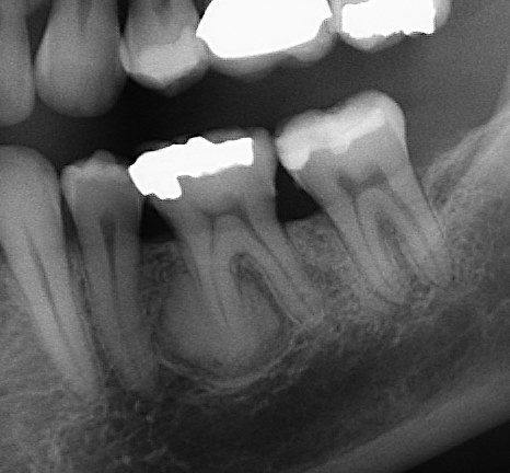 presented for evaluation of opaque lesion associated with mesial root of #19; Patient is asymptomatic, the lesion was found on routine radiographic examination; Tooth #19 is vital; no tenderness to palpation or percussion; There is on associated vestibular swelling;
The radiographic appearance of this lesion is diagnostic: Opaque lesion that is replacing the root of the tooth and is surrounded by a radiolucent ring.
Diagnosis: Cementoblastoma (true cementoma)
This is a rare, benign neoplasm of cementoblast origin. It is typically seen in the second and third decades of life, and there is no gender predilection. Mandibular teeth are involved more commonly than maxillary and posterior regions are involved more commonly than anterior. The tooth typically remains vital, there may be concurrent cortical expansion, and low-grade, intermittent pain may be present (not the case in this patient).
Microscopically the lesion consists of cementum-like material which is variably mineralized. The soft tissue component of the lesion is well vascularized, and contains numerous cementoblasts and cementoclasts.
Because of the association of the neoplasm with the root of the tooth this lesion cannot be removed without sacrificing the tooth. Removing the tooth requires removal of a large portion of bone to allow for the removal of the mass associated with the root. In this case, since patient was asymptomatic and the tooth is vital, radiographic monitoring of the lesion was chosen as the treatment approach.